Spiralling together: Into the Calcaneus
- Katie Davidson
- Apr 28
- 13 min read
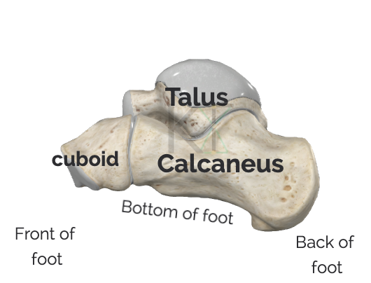
The calcaneus, is more commonly referred to as the heel bone, it is located at the back of the foot and will make contact with the ground when standing. The distinct shape of the bone is reflective of the joint surfaces located at the top and front of the bone, and the attachment points that shape the back and bottom of the bone.
Exploring the shape of the heel:
Back of the heel (posterior)The inner bottom part of the back of the heel makes the most contact with the floor when standing. The back of the bone contains the posterior tuberosity- which is the round edges at the very back of the bone. It is a major site for attachments. Tissues attaching to the back of the heel include but are not limited to the the calcaneal tendon, flexor retinaculum, and parts of the deltoid ligament..  | Bottom of the heel (plantar / sole)It is an important site for soft tissue attachments. including the plantar ligaments, aponeurosis It is an attachment point for flexor aponeurosis, the plantar muscles, and the plantar ligaments. Important structure in the longitudinal arches. Several blood arches cross the bottom of the heel and form pathways with the rest of the foot. 
|
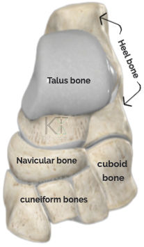
Front of the heel (anterior)It is a large saddle-like shape which makes contact with the talus and the cuboid bone, and contains the superior-medial projection called the talar shelf. The front of the bone is used as an attachment point for extensor digitorum brevis, and serves as a partition between the dorsal & plantar calcaneocuboidal ligaments.  | Top of the heel (superior / dorsal)Along the top inner heel, where the it meets the head of the talus, is the talar shelf. The head of the talus is considered the keystone to the medial long. arch. The talar shelf is a major attachment point for the ligaments.  |
Inner (medial) sideIt contains the medial talar shelf which is a projection along the top inner edge. It is a keystone in maintaining the high arch along the inner foot. It is a pathway for tendons, and NAVL moving between the foot and the leg.  | Outer (lateral) sideThere is a small arch along the outer edge of the heel. It contains the fibular trochlea that is a ridge located between the tendons of fibularis longus & brevis muscles, and NAVL. 
|
Joints of the Calcaneus

The heel bone has a lot of influence over the other bones of the feet. It forms joints with the talus bone and the cuboid bone, and it will have a more indirect influence over the ankle and the toes.
Sitting on top of the heel bone is the talus bone, together these 2 bones will carry the weight of the body and help send force towards the ground and towards the rest of the foot. These two bones have 3 distinct joint surfaces that connect to form the anatomical subtalar joint.
The heel bone will also have a joint surface along the outer front edge which forms part of the transverse joint. The transverse joint as a whole is actually a series of joints made up between several of the tarsal bones.

The main movement of these joints will be inversion and eversion which happens in the coronal plane, around an axis running from front to back of the foot.
The action of inversion brings the sole of the foot inwards while the outer edge of the foot rolls down towards the ground. The movement is made easier when pointing the big toe downwards.
Eversion brings the sole of the foot outwards, bringing weight onto the outer edge of the foot. This motion is made easier when the toes are lifted up towards the shin.
The subtalar joint
This joint can be described as the anatomical subtalar joint and the clinical subtalar joint.
The anatomical subtalar joint is a single synovial joint between the concave surface of the talus and the convex posterior articular facet of the calcaneus. Contains a weak joint capsule that is supported by several talo-calcaneal ligaments.
The clinical subtalar includes a part of the transverse joint. Creating a more functional joint within the foot because realistically it is very difficult to separate the joint between the tarsal bones.
Transverse talar joint
Formed by 2 separate joints, formed by 4 tarsal bones, moves in the coronal plane which cuts the body into front and back
Soft tissue related to the heel bone
Connective tissue (fascial connections)
Aponeurosis = thin sheet of fascia, it functions as attachment site for muscles, similar to tendons. Retinaculum = thickened pieces of fascia often found in areas with a lot of tendons (E.G feet and wrists). Retinacula keep tendons in the right stop and allow for proper movement of the tendon . |

Plantar aponeurosis → Thick, superficial, band of fascia located between the skin and the muscles. Travels from the back of the heel to the ball of the feet. It is an important structure for supporting the longitudinal arch.
Flexor retinaculum → broad strap extending from the medial calcaneus to the medial malleoli. Designed to hold in place the tendons of the flexor muscles and the tibial N&A


Superior fibular retinaculum → Extends from the lateral malleoli of the fibula to the lateral edge of the heel. It allows for the passage of the tendons of fibularis longus and brevis and the fibular artery and veins.
Muscles that attach to the calcaneus:
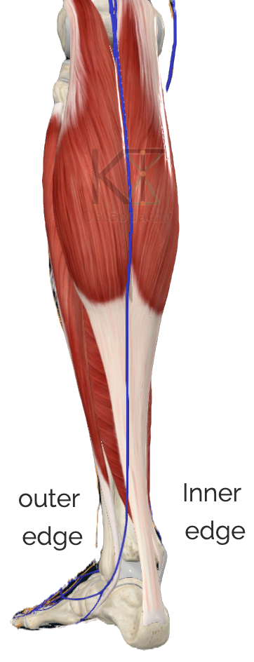
Superficial posterior compartment of the leg (calf):
 |  |  |
The superficial group of muscles at the back of the leg attach to the back of the heel bone. These muscles are the gastrocnemius muscle, the soleus muscle, and the plantaris muscle, all of which are innervated by the tibial nerve.
Their common movement is plantarflexion of the ankle that happens in the sagittal plane around an axis going from one side of the body to another (left to right) . This movement happens at the ankle joint making it an indirect joint of the heel.
The gastrocnemius and plantaris attach above the knee and also allow for knee flexion in the sagittal plane.
Based on muscle fiber type, the gastrocnemius muscle will be recruited during powerful movements such as jumping and running. The soleus muscle is used more for constant movements such as standing.
The plantaris muscle is rich in muscle spindles, which monitor stretching of the muscle and can create changes in the shape of the muscle if too much length is felt within the posterior compartment.
Many of the muscles of the leg will have tendons that cross over the calcaneus.
Muscles that move the toes:
 |  |  |
There are 5 muscles that attach to the heel and act on the toes. 1 on top of the heel and 4 on the bottom of the heel.
On the top of the bone is the extensor digitorum brevis. It will bring the toes that it attaches to up towards the shin (extension) which happens in the sagittal plane.
2 of the muscles on the bottom of the foot will help point the toes downwards: the quadratus plantae muscle and the flexor digitorum brevis muscle.67
The last 2 muscles move in the coronal plane around a horizontal axis and they are the abductor hallucis and abductor digiti minimi. They will be innervated by the medial and lateral plantar nerves.
Tendons that cross the calcaneus
 |  |  |
Tendons are rope like structures made from dense regular connective tissue that attaches muscles to bones, they are helpful in areas like the foot where there are many small bones that are recruited during movement.
From the deep part of the posterior compartment there will be 3 muscles that cross over the inner edge of the heel bone, passing deep to the flexor retinaculum. These muscles are the tibialis posterior, the flexor digitorum longus, and the flexor hallucis longus muscles . These muscles will plantarflex the ankle. The flexor hallucis longus will flex the big toe, and the flexor digitorum longus will flex the rest of the toes.

From the anterior compartment the tibialis anterior muscle will cross over the top of the foot. Not necessarily crossing over the calcaneus, but will move the subtalar joint through inversion along with tibialis posterior.
From the lateral compartment of the leg there are 2 muscles that cross over the outer edge of the heel bone, deep to the superior and inferior fibular retinacula. They are the fibularis longus and brevis muscles, which are the main contributors to eversion of the subtalar joints. In betwenn the tendons of fibularis longus and brevis, along the outer edge of the heel is the fibular trochlea.
Ligaments relating to the calcaneus
Ligaments are dense regular connective tissue that connect bones to other bones, they provide joint surfaces with
We will explore ligaments attaching to the plantar surface of the feet (sole), comparing the ligamentous structure on the medial versus lateral sides of the heel.
Bottom of the foot:

The spring ligament (plantar calcaneonavicular): It
attaches along the inner edge of the heel extending from the top to the bottom of the bone, it will then attach to the navicular bone. It supports the head of the talus, as its name suggests this ligament is important in take on force from the weight bearing talus.
Long plantar ligaments: It passes from the heel bone to the cuboid bone, and the metatarsal bones 2-5. The tendon of fibularis longus starts deep to this ligament along the lateral edge and becomes superficial as it crosses the bottom of the foot.
Short plantar ligament (Plantar calcaneocuboidal):
Extends from the the front part of bottom of the heel to the bottom of the cuboid bone. This is one of many tissues that help support the longitudinal arches of the feet.
Outer edge of the heel:
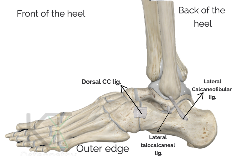
Calcaneofibular part of the lateral malleolar ligament: It attaches to the lateral malleolus of the fibula to the lateral surface of the heel. It stabilizes the ankle joint by limiting inversion. The tendons of fibularis long and brevis pass over top of the ligament.
Dorsal calcaneocuboid ligament: A thin broad ligament that attaches to the top outer edge of the calcaneus and the cuboid bones. It helps strengthen the joint capsule.
Lateral talocalcaneal ligament: It attaches to the bottom outer edge of the talus to the outer edge of the heel. It reinforces
Inner edge of the heel:

Tibiocalcaneal part of deltoid ligament (medial malleolar): A strong, fan-shaped ligament on the medial side of the ankle, attached to the talar shelf.
Medial talocalcaneal ligament: Attaches to the medial process passing obliquely to attach to the talar shelf on the calcaneus. It's a short flat ligament. Flexor hallucis long and tibial nerve passes superficial to the ligament.
Calcaneus’ role in maintaining the arches of the feet:

The foot is a complex structure in the human body,
when standing it must take on the weight of the body and distribute the force. That transfer of weight comes through the talus bone into the calcaneus and the rest of the bones in the feet. The heel and the sesamoid bones at the base of the big toe (circled) are large pointS of contact between the foot and the ground.
Between the heel and the toes are a series of arches formed by the bones, muscles, tendons, and ligaments. The arches run the length of the inner and outer edge of the foot and across the width of the foot. When standing the arches are relatively flat allowing for more contact between the foot and the ground.
Arches running the length of the foot:
Also known as longitudinal arches, there is a medial (inner) arch, and a lateral (outer) arch.
The inner arch is higher than the outer arch, the majority of the outer arch rests on the ground when standing. The inner part of the heel holds the head of the talus which marks the highest point of the inner arch. The tendons of tibialis posterior & anterior wrap around the inner heel and provide support to the inner arch. The tendon of fibularis longus wraps around the outer edge of the heel and crosses over to the inner edge of the foot. It supports the inner arch, and the transverse arch.
Arches running the width of the foot:
The arch the runs from side to side is known as the transverse arch, it is formed by the tarsal and metatarsal bones, the tendons of fibularis longus and tibialis posterior help maintain the arch.
The shape of the short bones, their joint surfaces, and supporting ligaments create the foundation for the bowstring like structure of soft tissues along the bottom of the foot. The muscles and tendons that attach to the sole of the foot provide contractile force that can change the shape of the curves.
Nerves, Arteries, Veins, Lymphatics relevant to calcaneus
NAVL for short. Nerves will innervate tissues. Arteries will supply the tissues Veins and lymphatic vessels will drain tissues. |
Arterial supply
Pathway to the popliteal artery Strats at the heart, travels through the major arteries in the thorax abdomen and pelvis. In front of the lumbar spine the common iliac artery divides into the internal and external iliac arteries.
The external iliac artery enter the thigh in front of the hips changing names to the femoral artery, as this artery descends in the thigh it moves backwards towards the back of the knee where it becomes the popliteal artery.
The popliteal artery is a good jumping off point to examine the blood supply to the heel. The posterior tibial artery is one of the terminal branches of the popliteal artery and is the main contribution to blood supply to the calcaneus.

The posterior tibial artery will descend in the back of the leg deep to the soleus muscle. Below the knee the posterior tibial artery will give off the fibular* branch (* sometimes comes off popliteal artery).
The fibular artery will descend along the outer edge of. the leg in between the fibularis longus and brevis muscles. It will then pass along the outer edge of the heel and give off a calcaneal and lateral malleolar branch.
Around the inner edge of the ankle the posterior tibial artery will give off a medial malleolar and calcaneal branch before dividing into its terminal branches: medial and lateral plantar arteries.

Blood flow in the foot is abundant, the arteries of the leg give off a lot of branches that form an anastomotic relationship. Which is essentially a mesh like network of blood vessels that provide alternate pathways for blood flow. Blood will always follow the path of least resistance, and this mesh like network of blood is a safeguard to protect blood flow incase of a blockage or injury that might impedes a blood vessel.
Venous drainage
Venous blood around the heel must make its way against gravity back towards the heart which is why the veins in the leg will contain valves so blood doesn't flow backwards.
There are two main venous pathways around the heel: a superficial route or deep pathway.
The deep pathway mirrors the main arterial pathway just in a reverse order and with a 2:1 ratio of veins to arteries. Oftentimes the doubled up veins will travel with the artery in between them.
Deep:
The venous plantar arch has drainage points into the medial and lateral plantar veins, creating a loop on the bottom of the foot deep to the flexor tendons. The medial and lateral plantar veins will joint together close to. the talar shelf of the heel and become the posterior tibial veins(2 of them). Along the outer heel there are the fibular veins (2 of them), which will travel with the fibular artery in between the,. The deep veins in the leg will drain into the popliteal vein.
there will be veins that connect the deep and superficial pathways which are called perforating veins.
Superficial:

Through the small and great saphenous veins, the small saphenous vein begins around the outer edge of the ankle. As it ascends it will move towards the midline of the calf, travelling in between the heads of gastrocnemius muscle and utilizing its contractile force to push blood upwards. The small saphenous vein will drain into the popliteal vein.
The great saphenous vein ascends along the inner edge of the ankle, it is considered the longest vein in the body. The great saphenous vein will travel the entire length of the leg and thigh to drain into the femoral vein close to the front of the hip.
Lymphatic drainage
Lymph vessels travel alongside the veins, so there will be superficial and deep vessels. the superficial vessels can be found along the small and great saphenous veins.
The vessels that follow the small saphenous vein will drain into popliteal lymph nodes. While the vessels following the great saphenous vein will drain close to the hip into the inguinal or iliac lymph nodes.
the deep lymph vessels accompany the deep veins and will drain into the popliteal lymph nodes.
Nervous innervation

The main nerve that will supply the heel and surrounding tissues is the sciatic nerve. It will travel from the lower lumbar spine and sacrum, through the glutes, down the the back of the thigh, knee and leg. Finally, the branches of the sciatic nerve will cross over the heel to supply the foot.

The sciatic nerve supplies to the back of the thigh and the majority of the leg and foot. This includes nerve supply to the muscles, joints, blood vessels, skin ...
Through it path in the pelvis and thigh the nerve fibers remain in one large bundle. At the lower end of the back of the thigh the sciatic nerve will divide into its main branches: The common fibular (peroneal) and the tibial nerve.
The tibial nerve is accompanied by the posterior tibial blood vessels creating a neurovascular bundle that passes deep to the soleus muscle and runs along the muscle bellies of the tendinous muscles of the leg.
The calf muscles and surrounding intermuscular septa protect the terminal branches of the sciatic nerve and blood vessels as they travel towards the heel. Nerve supply in the leg can come from the tibial nerve or one of the branches of the common fibular nerve. When it comes to the foot, the tibial nerve will provide motor and sensory innervation through its terminal branches: the medial and lateral plantar nerves.


Comments